
A mouse treated with surgery alone for glioblastoma survived, on average, 42 days afterward. A mouse given the same surgery plus a new light-activated nanoparticle treatment survived at a 100% rate through 60 days, the point where researchers stopped counting because every treated animal was still alive. That gap sits behind a new “double punch” nanoparticle platform developed jointly by researchers at the University of Technology Sydney, Harvard, and Henan University, published in Science Translational Medicine.
Glioblastoma earns its reputation as the deadliest common brain cancer through two compounding problems. Microscopic cancer cells spread into healthy surrounding tissue in threads too fine to see or fully remove, forcing surgeons to leave some cancer behind rather than risk damaging brain function nearby. What’s left afterward is largely untouchable by drugs and radiotherapy, since the blood-brain barrier blocks most treatment from reaching it in meaningful concentrations. Together, those two obstacles hold the five-year survival rate for glioblastoma at around 7%, a number that’s barely moved across decades of new treatment attempts.
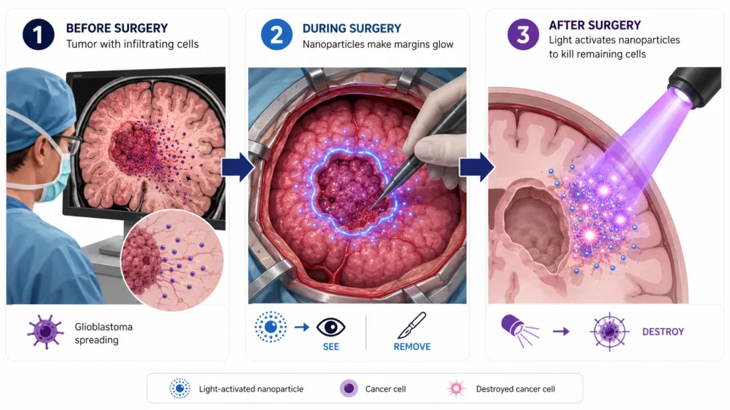
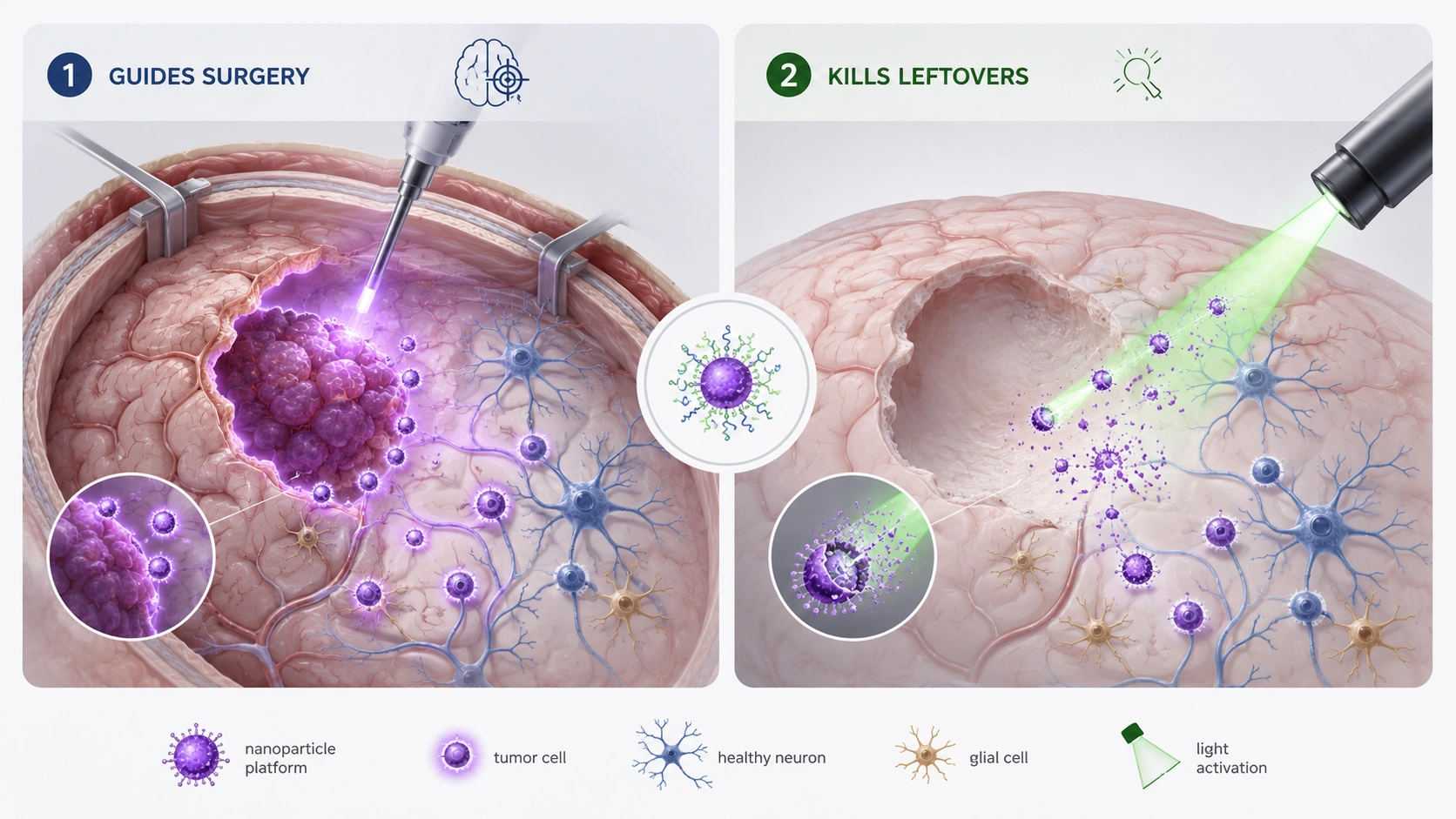
The nanoparticle platform tackles both problems with the same physical material rather than two separate tools. Chair professor of nanomedicine Bingyang Shi described the approach directly: we’ve engineered a single material that does two jobs in sequence. During surgery, the light-activated nanoparticles help surgeons see glioblastoma tissue with single-cell precision, sharpening the line between tumor and healthy brain in real time rather than relying on a surgeon’s eye alone. After surgery closes, the same material switches roles, activated by light to destroy whatever cancer cells remain in tissue the surgeon couldn’t safely remove. One tool, sequenced to do the seeing first and the killing second.
This isn’t the only nanoparticle strategy currently aimed at glioblastoma’s twin obstacles, and it’s worth knowing it sits inside a genuinely active field rather than standing alone. Separate teams have pursued different angles on the same basic problem: University of Virginia researchers have paired brain-penetrating nanoparticles with focused ultrasound to deliver gene-silencing molecules past the blood-brain barrier, work published through UVA’s own channels, while another group has tested sugar-coated nanoparticles carrying genetic instructions to restore a natural tumor-suppressing protein glioblastoma cells typically shut down, an approach detailed separately. A different research effort has focused on the brief window right after surgery when the blood-brain barrier itself becomes temporarily more permeable, repurposing already-approved nanoparticles to exploit that gap before it closes again. Each approach is chasing a version of the same goal from a different direction.
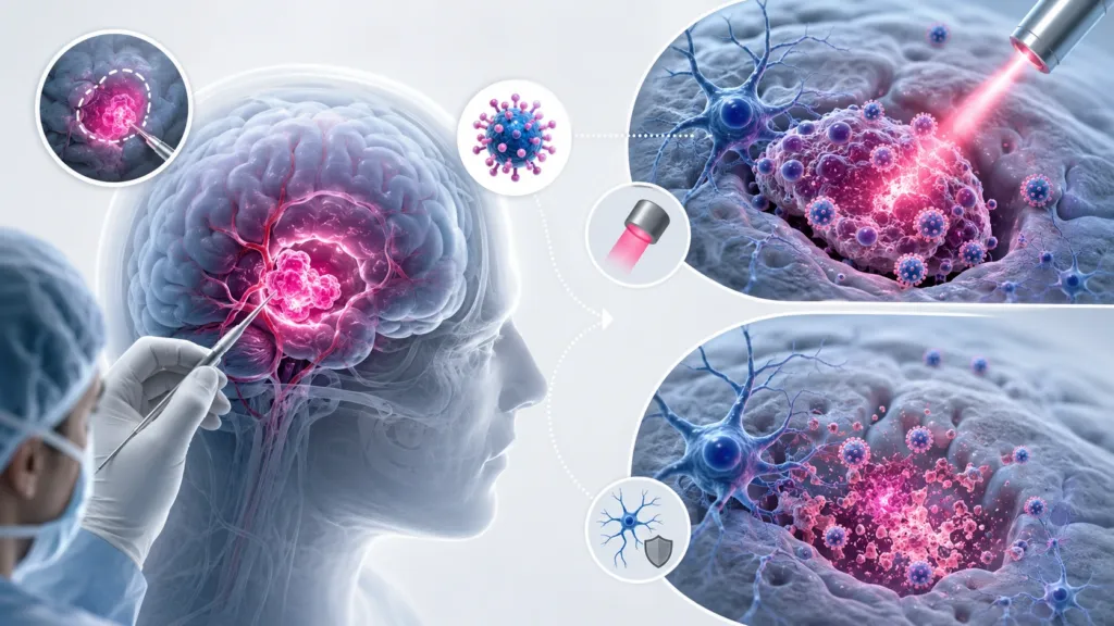
What distinguishes the UTS-Harvard-Henan platform is the dual function packed into one material, guidance during the operation and treatment after it, rather than requiring a separate visualization tool and a separate therapeutic delivered on their own timelines. That combination matters practically as much as scientifically. A surgical team already juggling imaging equipment, navigation systems, and the pressure of operating near brain regions that control speech, movement, or memory has less room for additional standalone tools, however promising each one is individually.
None of this is close to a human treatment yet. Every result described here comes from mouse models, and the jump from a mouse brain to a human one, particularly for something requiring precise light activation during live surgery, is a considerable engineering and regulatory distance still to travel. What the mouse data does show, a 100% survival rate against a 42-day baseline with no detectable neurological toxicity, is a large enough gap to justify the next phase of testing, the kind of gap that tends to justify moving quickly toward larger studies in a disease that has resisted meaningful survival gains for as long as glioblastoma has.